Thing need consider when find positioning in radiography?
When you looking for positioning in radiography, you must consider not only the quality but also price and customer reviews. But among hundreds of product with different price range, choosing suitable positioning in radiography is not an easy task. In this post, we show you how to find the right positioning in radiography along with our top-rated reviews. Please check out our suggestions to find the best positioning in radiography for you.
Best positioning in radiography


1. Clark's Positioning in Radiography 13E
Description
First published in 1939, Clarks Positioning in Radiography is the preeminent text on positioning technique for diagnostic radiographers.
Whilst retaining the clear and easy-to-follow structure of the previous edition, the thirteenth edition includes a number of changes and innovations in radiographic technique. The text has been extensively updated, including a new section on evaluating images, The 10-point plan, which is linked throughout to a listing of Essential image characteristics for each procedure. The section on digital imaging has been expanded not only to elaborate more extensively on the technology but to demonstrate its various clinical applications.
New sections also include imaging informatics and its role in the modern world of medical imaging, holistic approaches to patient care and discussion of the important aspect of the patient journey.
Students will also benefit from more detailed reference to positioning errors and how to avoid mistakes, as well as a greater emphasis on standard radiation protection measures and guidance on the most recent radiation dose reference levels for specific examinations.
Clarks Positioning in Radiography continues to be the definitive work on radiographic technique for all students on radiography courses, radiographers in practice and trainee radiologists.
2. Clark's Positioning in Radiography
Description
First published in 1939, Clark's Positioning in Radiography is the definitive text on patient positioning for the diagnostic radiography student and practitioner. This fully revised 12th edition ensures that the title retains its pre-eminence in the field, with hundreds of new positioning photographs and brand new explanatory line diagrams, a clearly defined and easy-to-follow structure, and international applicability. The book presents the essentials of radiographic techniques in as practical a way as possible, avoiding unnecessary technical complexity and ensuring that the student and practitioner can find quickly the information that they require regarding a particular position. The experienced author team, expanded for this edition, understand that there is no substitute for a good understanding of basic skills in patient positioning and an accurate knowledge of anatomy to ensure good radiographic practice. All the standard positioning is included in this single volume, accompanied by supplementary positions where relevant and illustrations of pathology where appropriate. Common errors in positioning are also included.3. Merrill's Atlas of Radiographic Positioning and Procedures: 3-Volume Set
Feature
MosbyDescription
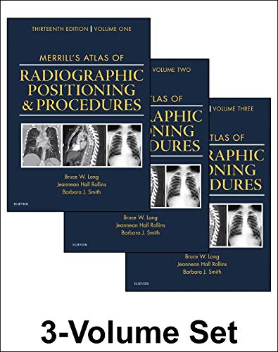
More than 400 projections make it easier to learn anatomy, properly position the patient, set exposures, and take high-quality radiographs! With Merrill's Atlas of Radiographic Positioning & Procedures, 13th Edition, you will develop the skills to produce clear radiographic images to help physicians make accurate diagnoses. It separates anatomy and positioning information by bone groups or organ systems using full-color illustrations to show anatomical anatomy, and CT scans and MRI images to help you learn cross-section anatomy. Written by radiologic imaging experts Bruce Long, Jeannean Hall Rollins, and Barbara Smith, Merrill's Atlas is not just the gold standard in radiographic positioning references, and the most widely used, but also an excellent review in preparing for ARRT and certification exams!
- UNIQUE! Collimation sizes and other key information are provided for each relevant projection.
- Comprehensive, full-color coverage of anatomy and positioning makes Merrill's Atlas the most in-depth text and reference available for radiography students and practitioners.
- Coverage of common and unique positioning procedures includes special chapters on trauma, surgical radiography, geriatrics/pediatrics, and bone densitometry, to help prepare you for the full scope of situations you will encounter.
- Numerous CT and MRI images enhance your comprehension of cross-sectional anatomy and help you prepare for the Registry examination.
- Bulleted lists provide clear instructions on how to correctly position the patient and body part when performing procedures.
- Summary tables provide quick access to projection overviews, guides to anatomy, pathology tables for bone groups and body systems, and exposure technique charts.
- Frequently performed projections are identified with a special icon to help you focus on what you need to know as an entry-level radiographer.
- NEW! Coverage of the latest advances in digital imaging also includes more digital radiographs with greater contrast resolution of pertinent anatomy.
- NEW positioning photos show current digital imaging equipment and technology.
- UPDATED coverage addresses contrast arthrography procedures, trauma radiography practices, plus current patient preparation, contrast media used, and the influence of digital technologies.
- UPDATED Pediatric Imaging chapter addresses care for the patient with autism, strategies for visit preparation, appropriate communication, and environmental considerations.
- UPDATED Mammography chapter reflects the evolution to digital mammography, as well as innovations in breast biopsy procedures.
- UPDATED Geriatric Radiography chapter describes how to care for the patient with Alzheimers Disease and other related conditions.
4. Handbook of Radiographic Positioning for Veterinary Technicians (Veterinary Technology)
Description
The Handbook of Radiographic Positioning for Veterinary Technicians is designed as a practical guide to positioning for radiographic studies in the small animal clinic. This concise reference presents a systematic approach to the positioning of canine, feline, and exotic animal patients for routine and special radiographic procedures. The primary focus is on providing visual aids of animals in position for radiographic procedures. The resulting radiograph produced is included for each radiographic position. A diagram of anatomical landmarks used in determining correct positioning is also included.5. Lange Radiographic Positioning Flashcards
Description
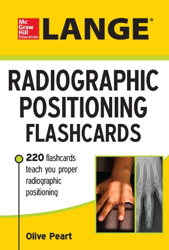
A comprehensive, carry-anywhere review of routine imaging procedures, projections, and positioning terminology
- Each two-sided card includes a high-quality photograph of correct patient positioning with details of the projection and the corresponding X-ray, technical information, and image evaluation criteria
- Most cards include a high-resolution radiographic image and photographs demonstrating each position/projection
- Great for use as a radiography procedures course review or as a clinical refresher prior to taking a patient's X-ray
6. Radiation Safety And Positioning Animals With Rope And Tape in Veterinary Radiography
Description
We get dozens of emails from technicians and practice owners about problems in their radiology department. Many of these problems are related to technician concerns about radiation safety, worries about exposure badge readings, increased exposure from unnecessary retakes, increased exposure from digital radiography, worries about pregnant employees, and technicians who just dont want to hand hold animals during radiography. This book addresses ALL of these issues and will help you take control of all radiology related issues in your practice. Every practice should have a book like this as part of their radiation safety training for new employees. This book is written in the most basic terms and even makes the painfully boring topic of radiation safety and entertaining an educational experience for the reader.7. Textbook of Radiographic Positioning and Related Anatomy
Feature
MosbyDescription
Focusing on one projection per page, Textbook of Radiographic Positioning and Related Anatomy, 8th Edition includes all of the positioning and projection information you need to know in a clear, bulleted format. Positioning photos, radiographs, and anatomical images, along with projection and positioning information, helpyou visualize anatomy and produce the most accurate images. With over 200 of the most commonly requested projections, this text includes all of the essential information for clinical practice.
- Lists and definitions of the most common pathologies likely to be encountered during specific procedures helps you understand the whole patient and produce radiographs that will make diagnosis easier for the physician.
- Labeled radiographs identify key radiographic anatomy and landmarks to help you determine if you have captured the correct diagnostic information on your images.
- Evaluation Criteria for each projection provide standards for evaluating the quality of each radiograph and help you produce the highest quality images.
- Clinical Indications sections explain why a projection is needed or what pathology is demonstrated to give you a better understanding of the reasoning behind each projection.
- Increased emphasis on digital radiography keeps you up to date with the most recent advances in technology.
- Completely updated content offers expanded coverage of important concepts such as, digital imaging systems, updated CT information and AART exam requirements.
- More CT procedures with related sectional images, especially for areas such as skull and facial bones, reflect the shift in the field from conventional radiography to CT.
- Updated art visually demonstrates the latest concepts and procedures with approximately 500 new positioning photos and 150 updated radiographic images.
- Additional critique images provide valuable experience analyzing images to prepare you to evaluate your own images in the practice environment.
- Updated Technique and Dose boxes reflect the higher kV now recommended for computed and digital radiography.
- Imaging Wisely program information from ASRT provides protocols to minimize radiation exposure during digital procedures.
- The latest standards for computed radiography and digital radiography (CR/DR) from the American Association of Physicists in Medicine ensures you are current with todays procedures and modalities.


Recent Comments